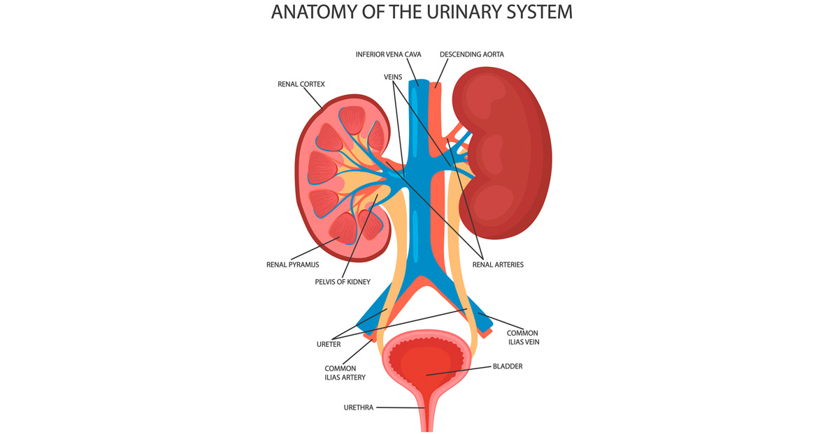

Uretero-pelvic junction (UPJ) obstruction occurs in the ureters, which are the tubes that connect the kidneys to the urinary bladder and transport urine produced by the kidneys. Conditions affecting the ureters may be treated with both endoscopic and laparoscopic procedures.
These uretero-pelvic junction (UPJ) procedures utilize minimally invasive techniques to treat UPJ conditions that require surgery. Advancements in technology have made it possible to use these treatment methods instead of traditional open surgery. Benefits of these methods for the patient include:
- Less pain
- Quicker procedure
- Less trauma to the body
- Shorter healing process
Endoscopic and Laparoscopic Treatment
Laparoscopy is a type of endoscopic surgery, which is simply a broad term for any type of minimally invasive surgery done with a small, flexible tube with a video camera and light on one end, called an endoscope. During laparoscopy, the surgeon makes incisions in the abdomen. Other endoscopies are done in other parts of the body and may not require incisions.
To treat ureter or uretero-pelvic junction conditions using an endoscopic method, a physician will use the endoscope to view the inside of the body and perform surgery without making a large six- to 12-inch incision typically done during open surgery.
During laparoscopic surgery, the surgeon will only make one to three incisions less than a half-inch long in the abdomen. He or she will insert tubing through the incisions, and the surgical instruments and laparoscope will be slid through the tubing. The surgical procedure will be performed on the ureter or uretero-pelvic junction with the help of a video monitor.

Ureteral Stone Treatment
Patients with kidney stones may receive endoscopic treatment to remove or break them up. To treat stones located in the ureter, the physician will perform a ureteroscopy. During this procedure, the physician inserts an endoscope into the urethra, through the bladder and into the ureter without making any incisions. If necessary, the stones will be broken up, and caught and removed with a small basket. A stent is usually temporarily left in the ureter to help keep it open and allow the kidney to drain.
Uretero-Pelvic Junction Obstruction
Uretero-pelvic junction obstruction, a condition more common in children than adults, may be treated with laparoscopic surgery. The condition is characterized by a total or partial block of the junction between the kidney and ureter, called the renal pelvis. This area can become enlarged when it is blocked because the ureter carries urine from the kidneys to the bladder. Left unaddressed, the result can be kidney damage.
Patients may need laparoscopic pyeloplasty to remove the blockage if the obstruction doesn’t correct itself or respond to antibiotics. During the procedure, the physician cuts the ureter from the renal pelvis, removes the obstruction, and reconnects the ureter to the renal pelvis. A temporary stent may be left in place to help drain the renal pelvis and kidney.
What to Expect During The Procedure
Treatment is typically performed under general anesthesia. Patients will need to stay in the hospital for one to two days after a laparoscopic pyeloplasty but may go home the same day after a ureteroscopy.
It’s normal to experience pain in the kidney area and when urinating for up to one week after surgery. Over-the-counter pain relievers may help patients manage the pain. Until the stent is removed, patients may have a feeling of fullness or an urgency to urinate. The body will adjust over time and symptoms will subside once the stent has been removed.
